Die AIM-Academy ist eine internationale Plattform für den Austausch von Wissen und Erfahrung. Unser oberstes Ziel es ist, Ergebnisse aus aktueller Forschung und Entwicklung in der dentalen Implantologie auszuwerten und darüber zu berichten. Der Hauptsitz ist in Lausanne. Die AIM Academy verbindet Zahnärzte, Chirurgen und Zahntechniker aus universitären und privaten Zentren zu einer internationalen implantologischen Gemeinschaft.
Die AIM-Academy wurde von Novodent im Juni 2018 in Lausanne/Schweiz gegründet. Gegenwärtig beläuft sich die Mitgliederzahl auf 1.600 mit stark steigender Tendenz. Der erste und zweite internationale Jahreskongress im November 2018 (Antalya) und im Oktober 2019 (Kiew) konnten jeweils fast 1.000 Teilnehmer begrüßen.
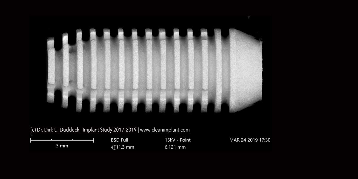
Die AIM-Academy unterstützt präklinische und klinische Studien, um evidenzbasierte Daten zu implantologischen Fragestellungen zu gewinnen.
Wir schaffen ein weltumspannendes soziales und fachliches Netzwerk für dentale Implantologie. Die AIM-Academy ist mittlerweile in 15 Mitgliedsstaaten vertreten, wo in regionalen Arbeitsgruppen auf nationaler Ebene regelmäßig Konferenzen stattfinden.
Im Rahmen eines jährlichen, internationalen Symposiums mit hochkarätigen Referenten wird unser wissenschaftlicher Anspruch untermauert.
Besuchen Sie unsere Kurse in einem unserer ImplantConcept Center und nehmen Sie an unseren Events an ausgewählten Orten der Welt teil.
Sie sind herzlich eingeladen, für unsere Jahreskongresse Ihre Präsentationen dem wissenschaftlichen Board einzureichen.
Die AIM-Academy schenkt Ihnen den persönlichen Kontakt zu Kollegen aus fremden Ländern und Kontinenten. Lernen Sie neue Aspekte der dentalen Implantologie kennen.
Werden Sie Teil unserer Vision.
Jetzt Abstract einreichen…
Unsere aktuellen Veranstaltungen
25.10.2019 - ImplantConcept Center Bochum
Element1 TEST
Element2 TEST
18.-20.10.2018 2. Internationaler Jahreskongress AIM Academy Kiew
Future Trends in Oral Implantology
In der atemberaubenden Kulisse des Oktober Palastes in Kiew findet der 2. Internationale Jahreshauptkongress der AIM Academy statt. Renommierte Referenten aus 15 Nationen werden über die zukünftigen Trends in der oralen Implantologie berichten.
Buchen Sie jetzt!
15.11.2019 - ImplantConcept Center Zürich
This is a text block. Click the edit button to change this text.


Jetzt Veranstaltung buchen…
Unsere bisherigen Veranstaltungen
16.-18.10.2018 1. Internationaler Jahreskongress AIM Academy Antalya
Contemporary Trends in Dental Implantology
Die AIM Academy konnte zum ersten Jahreskongress fast 1000 Mitglieder aus 28 Nationen begrüßen.
25.10.2019 - ImplantConcept Center Bochum
Element1 TEST
Element2 TEST
15.11.2019 - ImplantConcept Center Zürich
This is a text block. Click the edit button to change this text.
Unsere Publikationen
20 June 2018 - Journal of Clinical Periodontology
Europerio 9, Amsterdam 20-23 June 2018, Journal of Clinical Periodontology 2018;45(Supplement s19):343-344.
Maxillary sinus floor elevation using transalveolar approach and simultaneous extra‐short implant placement: a prospective clinical study
Akcali, A.1, Gurlek, O.2, Nizam N2.
Author information
1 Department of Endodontics, School of Dentistry, Ege University, Witten, Germany.
2 Department of Periodontology, School of Dentistry, Ege University, İzmir, Turkey.
Abstract
BACKGROUND AND AIM:
To evaluate clinical and radiographic outcomes of extra‐short implants placed either in combination with transalveolar maxillary sinus floor elevation or alone.
MATERIALS AND METHODS:
Twenty‐four systemically and periodontally healthy, non‐smoker patients (13 females and 11 males, mean age ± SD; 52.83 ± 10.02) having at least one missing tooth in posterior maxillary area were included to the study. In the test group (21 implants, residual alveolar bone height (RBH) 2–5 mm) transalveolar approach with grafting material and simultaneous implant placement was performed while in the control group (24 implants, RBH >5 mm) implants were placed without any augmentation. The diameter and the length of the implants were ranged as 4.5–6.0 and 5.0–6.0 respectively. Second stage surgery was performed at third month and then implants were received single unit porcelain fused to metal crowns. Outcome measures including clinical and radiographic evaluations (periapical x‐rays) were recorded at baseline (delivery of the prosthesis), 6‐ and 12‐months after implant loading. Crestal bone levels (CBL), crown to implant ratios and transalveolarly augmented sinus heights were calculated digitally.
RESULTS:
Postoperative complications were not observed in both groups. CBL were similar among test and control groups during all time points of evaluation (p > 0.05). CBL changes at 6‐months (test: 0.09 ± 0.36 mm, control: 0.12 ± 0.38 mm) (p = 0.77) and 12‐months (test: 0.10 ± 0.30 mm, control: 0.17 ± 0.36 mm) (p = 0.51) after implant loading was also similar between groups. The crown to implant ratio and height of the augmented sinus did not correlate with the CBL.
CONCLUSION:
Extra‐short implant placement with transcrestal approach in sites with challenging RBHs and non‐splinted restoration of the placed implants might be promising treatment option. After 1‐year implant loading period success rates of the implants are not influenced by limited RBH as well as height of the elevated sinus.
This article is protected by copyright. All rights reserved.
29.11.-30.11.2018 EAO Wien
EAO 28, Vienna, Clinical Oral Implants Research, Volume 29, Issue S17.
4 mm bone level implants in the treatment of single tooth‐premolar sites – a case series. implants in the prosthetic rehabilitation of the posterior maxilla.
Avci A.1, Nizam N1.
Author information
- 1 Department of Periodontology, School of Dentistry, Ege University, İzmir, Turkey.
Abstract
AIM:
The aim of the present case series was to evaluate the 1-year clinical results of 4mm long bone level implants used to rehabilitate single premolar gap in mandible.
MATERIALS AND METHODS:
Six systemically healthy, non-smoker patients (3 male, 3 female, age range 48-62) with a single missing premolar tooth in the mandible were included in the study. A two stage surgical protocol was followed and the patients received a total of 6 implants, 4.0mm in length and ø 5.0 or 5.5mm (i-system, Novodent S.A., Switzerland). The implants were uncovered at 3rd. month and a single porcelain fused to metal restoration was fabricated thereafter. Radiographic evaluations were performed at baseline and 6-, 12-months after loading. Measurements were done by a digital image analysis system.
RESULTS:
The implants osseointegrated uneventfully with no sign of postoperative complications. All the implants remained in function during the evaluation period. Crystal bone loss was 0.10 ± 0.19mm and 0.19 ± 0.36mm at 6 and 12 months respectively.
CONCLUSION:
With the limits of the present short term clinical case series, it may be suggested that 4mm short implants may be regarded as a promising treatment option in the rehabilitation of single premolar gaps in the mandible.
This article is protected by copyright. All rights reserved.
29.07.2019 - Australian Dental Journal
Aust Dent J. 2019 Jul 29. doi: 10.1111/adj.12711. [Epub ahead of print]
Extra short implants in the prosthetic rehabilitation of the posterior maxilla.
Gürlek Ö1, Kaval ME2, Buduneli N1, Nizam N1.
Author information
- 1 Department of Periodontology, School of Dentistry, Ege University, İzmir, Turkey.
- 2 Department of Endodontics, School of Dentistry, Ege University, İzmir, Turkey.
Abstract
AIM:
To compare clinical outcomes of „extra short“ and regular bone level implants in the posterior maxilla for 12-months after loading.
MATERIALS AND METHODS:
Twenty-three systemically healthy, non-smoking patients received 30 extra short, 24 regular bone level implants. Acrylic stents were fabricated for each patient for correct implant positioning. Implant lengths were 4-6 mm in the test, 8 mm/10 mm in the control group. Radiographic evaluation was performed at baseline, 6-, and 12-months after loading. Crestal bone level (CBL), CBL change (CBLC), true crown length (TCL), implant/crown ratio (ICR) and residual bone height (RBH) below maxillary sinus floor were calculated digitally. Data were tested statistically.
RESULTS:
RBH was significantly lower and TCL, ICR were higher in the test than the control group (p<0.0001). CBL measurements at baseline were 0.19±0.18 mm and 0.31±0.37 mm and at the 12-months 0.24±0.24 mm and 0.41±0.31 mm, respectively in the test and control groups. CBL values at 12-months were significantly lower in the test than the control group (p<0.05). CBLCs were similar at all times (p>0.05). No correlation was found between the CBLC and implant/prosthetic parameters.
CONCLUSION:
Extra short and regular implants may provide similar clinical outcomes in prosthetic rehabilitation of atrophic maxilla, during 12-months follow-up. MeSH Key words: dental implants; maxilla; osseointegration; radiography.
This article is protected by copyright. All rights reserved.
25.-27. April 2019 - International Symposium Osteology in Barcelona
International Symposium Osteology, Barcelona, Spain, 25-27 April 2019.
Sinus floor elevation utilizing transalveolar approach with either synthetic bone substitutes or autografts followed by simultaneous short implant placement: a 1-year follow-up.
Author information
1 Department of Endodontics, School of Dentistry, Ege University, Witten, Germany.
2 Department of Periodontology, School of Dentistry, Ege University, İzmir, Turkey.
Abstract
AIM:
To compare clinical outcomes of „extra short“ and regular bone level implants in the posterior maxilla for 12-months after loading.
MATERIALS AND METHODS:
Twenty-three systemically healthy, non-smoking patients received 30 extra short, 24 regular bone level implants. Acrylic stents were fabricated for each patient for correct implant positioning. Implant lengths were 4-6 mm in the test, 8 mm/10 mm in the control group. Radiographic evaluation was performed at baseline, 6-, and 12-months after loading. Crestal bone level (CBL), CBL change (CBLC), true crown length (TCL), implant/crown ratio (ICR) and residual bone height (RBH) below maxillary sinus floor were calculated digitally. Data were tested statistically.
RESULTS:
RBH was significantly lower and TCL, ICR were higher in the test than the control group (p<0.0001). CBL measurements at baseline were 0.19±0.18 mm and 0.31±0.37 mm and at the 12-months 0.24±0.24 mm and 0.41±0.31 mm, respectively in the test and control groups. CBL values at 12-months were significantly lower in the test than the control group (p<0.05). CBLCs were similar at all times (p>0.05). No correlation was found between the CBLC and implant/prosthetic parameters.
CONCLUSION:
Extra short and regular implants may provide similar clinical outcomes in prosthetic rehabilitation of atrophic maxilla, during 12-months follow-up. MeSH Key words: dental implants; maxilla; osseointegration; radiography.
This article is protected by copyright. All rights reserved.
März/April 2020 - Journal of Oral and Maxillofacial Implants
Int J Oral Maxillofac Implants. 2020 Mar/Apr;35(2):415-422. doi: 10.11607/jomi.7950.
Extra-Short Implants With Osteotome Sinus Floor Elevation: A Prospective Clinical Study
Nizam N1, Gürlek Ö1, Kaval ME2.
Author information
- 1 Department of Periodontology, School of Dentistry, Ege University, İzmir, Turkey.
- 2 Department of Endodontics, School of Dentistry, Ege University, İzmir, Turkey.
Abstract
Purpose: The aim of this study was to assess the radiographic and clinical outcomes of extra-short implants either alone or in conjunction with osteotome sinus floor elevation and to compare these with regular-sized implants in the posterior atrophic maxilla.
Materials and methods: Systemically healthy, nonsmoker individuals having at least one tooth gap in the posterior maxilla were included in the study. When the residual bone height was < 4 mm, an extra-short implant (4 to 6 mm) in conjunction with osteotome sinus floor elevation was placed; when the residual bone height was between 4 and 7 mm, an extra-short implant alone was placed; and when it was ≥ 8 mm, a regular implant (8 to 10 mm) was placed. The implants were uncovered at 4 months, and porcelain-fused-to-metal crowns were fabricated. Crestal bone level, change in the crestal bone level, crown-to-implant ratio, and residual bone height were measured at baseline and 6 and 18 months postloading.
Results: Thirty patients (15 men, 15 women, age range: 30 to 73 years) received 80 implants. One implant in the extra-short implant (n = 27 implants) and regular implant (n = 24 implants) groups and two implants in the extra-short implant with osteotome sinus floor elevation group (n = 29 implants) failed before loading. Crestal bone level was significantly higher in the regular implant group compared with the extra-short implant with osteotome sinus floor elevation group at 18 months (P < .028). Crestal bone level change between 6 and 18 months was significantly lower in the extra-short implant + osteotome sinus floor elevation group compared with the regular implant group (P = .003). There was no correlation between the crestal bone level, crestal bone level change, and prosthetic and implant characteristics (P > .05).
Conclusion: Extra-short implants placed either in native bone or in conjunction with osteotome sinus floor elevation may provide similar clinical and radiographic outcomes that are comparable to those obtained with regular implants. Both extra-short implant placement methods can be promising noninvasive treatment options for the posterior maxilla, and implant dimension, crown length, crown-to-implant ratio, and residual bone height may not affect the crestal bone level change, at least in the short term.